Skip Navigation
Skip to contents

JMD : Journal of Movement Disorders

OPEN ACCESS
SEARCH
Search

Articles

Page Path
HOME > J Mov Disord > Volume 17(1); 2024 > Article
Letter to the editor
A Novel Pathogenic PSEN1 Variant in a Patient With Dystonia-Parkinsonism Without Dementia
Maria Chiara Malaguti1corresp_iconorcid, Alessio Di Fonzo2orcid, Chiara Longo1,3orcid, Raffaella Di Giacopo4orcid, Costanza Papagno5orcid, Davide Donner6,7orcid, Umberto Rozzanigo8orcid, Edoardo Monfrini2,9orcid
Journal of Movement Disorders 2024;17(1):102-105.
DOI: https://doi.org/10.14802/jmd.23125
Published online: September 14, 2023

1Department of Neurology, Santa Chiara Hospital, APSS, Trento, Italy

2Department of Neurology, Foundation Istituti di Ricovero e Cura a Carattere Scientifico Ca’ Granda Ospedale Maggiore Policlinico, Milan, Italy

3Department of Psychology, University of Milano-Bicocca, Milan, Italy

4Department of Neurology, Rovereto Hospital, APSS, Rovereto, Italy

5Center for Mind/Brain Sciences (CIMeC), University of Trento, Rovereto, Italy

6Department of Nuclear Medicine, Santa Chiara Hospital, APSS, Trento, Italy

7Department of Medical and Surgical Sciences, University of Bologna, Bologna, Italy

8Department of Diagnostic Imaging, Santa Chiara Hospital, APSS, Trento, Italy

9Department of Pathophysiology and Transplantation, Neuroscience Section, Dino Ferrari Center, University of Milan, Milan, Italy

Corresponding author: Maria Chiara Malaguti, MD Department of Neurology, Santa Chiara Hospital, APSS, Largo Medaglie d’Oro, 9, Trento 38122, Italy / Tel: +39-0461-903281 / Fax: +39-0461-902732 / E-mail: mariachiara.malaguti@apss.tn.it
• Received: June 30, 2023   • Revised: August 10, 2023   • Accepted: September 13, 2023

Copyright © 2024 The Korean Movement Disorder Society

This is an Open Access article distributed under the terms of the Creative Commons Attribution Non-Commercial License (http://creativecommons.org/licenses/by-nc/4.0/) which permits unrestricted non-commercial use, distribution, and reproduction in any medium, provided the original work is properly cited.

prev next
  • 1,324 Views
  • 71 Download
Dear Editor,
The PSEN1 gene is located on chromosome 14 and encodes the presenilin 1 protein, which is a key component of the γsecretase complex that is involved in the cleavage of amyloid precursor protein (APP) in amyloid beta (Aβ) peptides [1]. Recent works have suggested that Aβ peptides may also play a role in the pathophysiology of motor symptoms in Alzheimer’s disease (AD) patients carrying PSEN1 mutations (PSEN1-AD) through accumulation in the striatum [2]. Aβ peptides may disrupt the function of the basal ganglia, possibly leading to the development of extrapyramidal symptoms [3]. In addition, PSEN1 mutations can be associated with the accumulation of other proteins, such as alpha-synuclein and tau, which have also been linked with motor signs [4]. Parkinsonism, ataxia, and spasticity are the most frequently described motor symptoms in PSEN1-AD patients [5]. Motor impairment has been reported in the early stages of the disease and may even precede cognitive decline in a small subset of patients [5]. Few cases have been reported of PSEN1 mutation carriers with parkinsonism as an isolated presenting feature without dementia or significant cognitive decline over time [6-8]. Here, we report a male patient with adult-onset dystonia-parkinsonism with positive AD biomarkers carrying a novel PSEN1 frameshift variant.
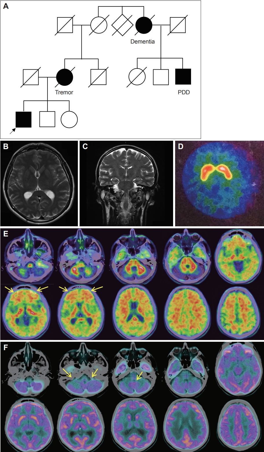
A 52-year-old man came to our observation for painful sustained muscular contractions, cramps, and subjective mild attention deficit. Neurological examination revealed lower limb dystonia and symmetric rigidity causing gait impairment, rest and postural right-hand tremor, mild spontaneous and sensoryinduced myoclonus, global and symmetric bradykinesia, mild cerebellar dysarthria and fluctuating diplopia (Supplementary Video 1 in the online-only Data Supplement). He had a positive family history of neurological diseases on the maternal side of the family with a hereditary pattern compatible with autosomal dominant inheritance (i.e., head tremor in his mother and Parkinson’s disease evolving to dementia in a second-degree cousin and dementia in a great aunt, both from the maternal side) (Figure 1A).
Blood test results were normal, including systemic autoimmunity, neurological paraneoplastic antibodies, and ceruloplasmin. Brain magnetic resonance imaging (MRI) was unremarkable (Figure 1B, C), while single photon emission computed tomography (SPECT) with ioflupane (123I) showed a moderate reduction in radiotracer uptake in the striatum with a slight right-side prevalence (Figure 1D). In-depth cognitive assessment revealed substantially preserved cognition with selective difficulty in visuospatial short-term memory and learning verbal material in the absence of executive deficits (Mini Mental State Evaluation [MMSE] = 29/30, Clinical Dementia Rating scale [CDR] = 0.5).
He started levodopa and pramipexole with partial motor benefit (highest levodopa equivalent daily dose [LEDD] = 650 mg). The discrepancy between OFF Unified Parkinson’s Disease Rating Scale III (UPDRS-III) (39) and ON UPDRS-III (30) was low. Pramipexole caused impulse control disorders (ICDs), namely, hyperphagia, compulsive shopping, and pathological gambling. This symptomatology subsided after pramipexole discontinuation.
Seven years after disease onset, at 59 years old, the patient presented with abrupt-onset status dystonicus requiring hospitalization. No clear trigger was identified. Neurological examination showed generalized dystonia, stiffness, and generalized myoclonic jerks, in addition to the parkinsonian phenotype. Status dystonicus was successfully treated with levodopa, oral clonazepam, and high doses of intravenous diazepam.
A clinical diagnosis of atypical parkinsonism associated with dystonia and myoclonus of suspected hereditary origin was made. After obtaining written informed consent, the patient’s DNA was tested with whole-exome sequencing (WES). A virtual panel including genes associated with parkinsonism, dystonia, and dementia was used. A novel heterozygous PSEN1 variant was identified (c.121_124dup, p.Arg42Glnfs*8) and validated by Sanger sequencing. WES did not reveal any other candidate variants. The PSEN1 c.121_124dup (p.Arg42Glnfs*8) is a novel frameshift variant predicted to lead to early protein truncation. Since loss of function is the known disease mechanism, this variant was classified as likely pathogenic (American College of Medical Genetics and Genomics criteria: PVS1 and PM2). Affected relatives were not available for genetic testing.
The identification of this likely pathogenic PSEN1 variant prompted us to reverse-phenotype the patient, looking for AD phenotypes and biomarkers. Cerebrospinal fluid (CSF) analysis showed positive AD biomarkers (total Tau 509 pg/mL [normal values: 146–410 pg/mL], P-Tau 79.1 pg/mL [normal value: 21.5–59.0 pg/mL], Aβ42 662 pg/mL [normal value: 725–1,777 pg/mL], and Aβ42/Aβ40 ratio 0.045 [normal value: 0.068–0.115]). 18F-Flutemetamol positron emission tomography-computed tomography (PET-CT) revealed symmetrical accumulation of Aβ in the cortical layer of the frontal cortex (Figure 1E). Finally, 18-Fluorodeoxyglucose (FDG)-PET showed symmetrical cerebellar hypometabolism (Figure 1F). Fifteen years after clinical onset, neurological examination showed substantial motor stability of the dystonic-parkinsonian phenotype and a preserved cognitive profile with a mild impairment in visuospatial short-term memory in the absence of long-term memory or significant executive function deficits (MMSE = 28/30; CDR = 0.5).
PSEN1 mutations are typically associated with familial earlyonset AD, but atypical phenotypes have been reported, and evidence is accumulating regarding the possibility of a PSEN1-related parkinsonian phenotype. Indeed, likely pathogenic PSEN1 variants have been identified in a few patients with early-onset levodopa-responsive parkinsonism (Supplemental Table 1 in the online-only Data Supplement). For example, Carecchio et al. [6] described a patient with early-onset dystonia-parkinsonism with cognitive impairment carrying a de novo PSEN1 mutation. AD biomarkers were not available in this case. Gatto et al. [7] described a woman with early-onset levodopa-responsive parkinsonism and mild non-amnestic cognitive deficit. Classic AD biomarkers (18FDG-PET, 18F-flutemetamol PET-CT, and CSF biomarkers) were inconclusive [7]. Finally, Chen et al. [8] described a patient with levodopa-responsive parkinsonism, mild cognitive impairment, and positive PiB-PET, harboring a de novo PSEN1 variant. Data from previous case reports concerning cognitive functions and AD biomarker findings are not consistent. Our patient manifested with dystonia-parkinsonism in the absence of typical cognitive features of AD. However, CSF analysis and 18F-flutemetamol PET-CT revealed the presence of AD biomarkers, suggesting an underlying AD pathology.
The PSEN1-related motor phenotype appears to be characterized by motor onset with rigidakinetic symptoms (not necessarily lateralized) that can progress to dystonic, pseudobulbar and/or pyramidal signs. The reduced response to levodopa and atypical phenotypic features may be due to the different pattern of neuropathological damage associated with PSEN1 mutation in comparison to typical Parkinson’s disease.
No clear genotype-phenotype correlations are possible to date, since reported PSEN1-related cases with a parkinsonian phenotype are extremely rare and the associated mutations do not localize in a specific protein domain. Large clinical, genetic, and neuropathological studies are needed to elucidate the relationships between PSEN1 mutations, AD pathology, and extrapyramidal phenotypes.
In conclusion, we reported a case of a PSEN1-related dystonic and parkinsonian phenotype harboring a novel truncating PSEN1 mutation. The clinical phenotype of this patient was dominated by disorders of movement, including dystonia, parkinsonism with partial levodopa responsiveness, and myoclonus in association with mild and non-progressive cognitive deficits. This case highlights the need to further investigate PSEN1 mutations as a likely genetic cause of adult-onset dystonia-parkinsonism even in the absence of dementia.
The online-only Data Supplement is available with this article at https://doi.org/10.14802/jmd.23125.

Video 1.

The video shows the patient’s gait impairment caused by lower limb dystonia and stiffness, right upper and lower limb tremor at rest, bilateral bradykinesia with finger and foot tapping with slight mirror movements in the right hand, bilateral striatal toes and a slightly abnormal pull test with mild postural instability.

Supplemental Table 1.

Motor, cognitive and imaging findings of clinical cases of PSEN1-related parkinsonian phenotype
jmd-23125-Supplementary-Table-1.pdf

Ethics Statement

The authors confirm that the approval of an Institutional Review Board (IRB) was not required for this work. Informed consent was obtained from the patient for genetic testing and case report. The authors certify that this article complies with the Principles of Ethical Publishing of Journal of Movement Disorders and declare that they acted in accordance with ethical standards laid down in the 1964 Declaration of Helsinki and its later amendments.

Conflicts of Interest

The authors have no financial conflicts of interest.

Funding Statement

None

Author contributions

Conceptualization: Maria Chiara Malaguti, Alessio Di Fonzo, Edoardo Monfrini. Project administration: Maria Chiara Malaguti, Alessio Di Fonzo, Edoardo Monfrini. Data curation: Maria Chiara Malaguti, Alessio Di Fonzo, Chiara Longo, Edoardo Monfrini. Investigation: all authors. Methodology: Maria Chiara Malaguti, Alessio Di Fonzo, Edoardo Monfrini. Visualization: Maria Chiara Malaguti, Alessio Di Fonzo, Edoardo Monfrini. Writing—original draft: Maria Chiara Malaguti, Chiara Longo, Alessio Di Fonzo, Edoardo Monfrini. Writing—review & critique: all authors.

Figure 1.
Family pedigree and neuroimaging. A: Family pedigree. Fully black symbols indicate individuals reported by the patient (arrow) and relatives to be neurologically affected. B, C: Normal brain MRI (T2 axial and coronal sections). D: SPECT with ioflupane (123I) shows a moderate reduction of radiotracer uptake in the striatum with a slight right-side prevalence. E: 18F-Flutemetamol PET-CT displays symmetrical accumulation of Aβ in the cortical layer of the frontal cortex (arrows). F: 18-Fluorodeoxyglucose (FDG)-PET shows symmetrical cerebellar hypometabolism and relative higher uptake in the vermis (arrows). PDD, Parkinson’s disease dementia; MRI, magnetic resonance imaging; SPECT, single photon emission computed tomography; PET, positron emission tomography; CT, computed tomography.
jmd-23125f1.jpg
  • 1. Jayne T, Newman M, Verdile G, Sutherland G, Münch G, Musgrave I, et al. Evidence for and against a pathogenic role of reduced γ-secretase activity in familial Alzheimer’s disease. J Alzheimers Dis 2016;52:781–799.ArticlePubMed
  • 2. Takao M, Ghetti B, Hayakawa I, Ikeda E, Fukuuchi Y, Miravalle L, et al. A novel mutation (G217D) in the Presenilin 1 gene (PSEN1) in a Japanese family: presenile dementia and parkinsonism are associated with cotton wool plaques in the cortex and striatum. Acta Neuropathol 2002;104:155–170.ArticlePubMedPDF
  • 3. Winkel I, Ermann N, Żelwetro A, Sambor B, Mroczko B, Kornhuber J, et al. Cerebrospinal fluid α synuclein concentrations in patients with positive AD biomarkers and extrapyramidal symptoms. J Neural Transm (Vienna) 2021;128:817–825.ArticlePubMedPMCPDF
  • 4. Bagaria J, Bagyinszky E, An SSA. Genetics, functions, and clinical impact of presenilin-1 (PSEN1) gene. Int J Mol Sci 2022;23:10970.ArticlePubMedPMC
  • 5. Zhang S, Lei C, Liu P, Zhang M, Tao W, Liu H, et al. Association between variant amyloid deposits and motor deficits in FAD-associated presenilin-1 mutations: a systematic review. Neurosci Biobehav Rev 2015;56:180–192.ArticlePubMed
  • 6. Carecchio M, Picillo M, Valletta L, Elia AE, Haack TB, Cozzolino A, et al. Rare causes of early-onset dystonia-parkinsonism with cognitive impairment: a de novo PSEN-1 mutation. Neurogenetics 2017;18:175–178.ArticlePubMedPDF
  • 7. Gatto EM, Rojas GJ, Nemirovsky SI, Da Prat G, Persi G, Cesarini M, et al. A novel mutation in PSEN1 (p.Arg41Ser) in an Argentinian woman with early onset parkinsonism. Parkinsonism Relat Disord 2020;77:21–25.ArticlePubMed
  • 8. Chen Y, Liu P, Xie F, Wang B, Lin Z, Luo W. A heterozygous de novo PSEN1 mutation in a patient with early-onset parkinsonism. Neurol Sci 2022;43:1405–1409.ArticlePubMedPDF

Figure & Data

References

    Citations

    Citations to this article as recorded by  

      Comments on this article

      Add a comment
      Figure
      • 0
      Related articles
      A Novel Pathogenic PSEN1 Variant in a Patient With Dystonia-Parkinsonism Without Dementia
      Image
      Figure 1. Family pedigree and neuroimaging. A: Family pedigree. Fully black symbols indicate individuals reported by the patient (arrow) and relatives to be neurologically affected. B, C: Normal brain MRI (T2 axial and coronal sections). D: SPECT with ioflupane (123I) shows a moderate reduction of radiotracer uptake in the striatum with a slight right-side prevalence. E: 18F-Flutemetamol PET-CT displays symmetrical accumulation of Aβ in the cortical layer of the frontal cortex (arrows). F: 18-Fluorodeoxyglucose (FDG)-PET shows symmetrical cerebellar hypometabolism and relative higher uptake in the vermis (arrows). PDD, Parkinson’s disease dementia; MRI, magnetic resonance imaging; SPECT, single photon emission computed tomography; PET, positron emission tomography; CT, computed tomography.
      A Novel Pathogenic PSEN1 Variant in a Patient With Dystonia-Parkinsonism Without Dementia

      JMD : Journal of Movement Disorders Twitter
      Close layer
      TOP