Articles
- Page Path
- HOME > J Mov Disord > Volume 3(2); 2010 > Article
-
Case Report
A Case of Action-Induced Clonus that Mimicked Action Tremors and was Associated with Cervical Schwannoma - Young-Hee Sung, Ki-Hyung Park, Yeung-Bae Lee, Hyeon-Mi Park, Dong-Jin Shin
-
Journal of Movement Disorders 2010;3(2):48-50.
DOI: https://doi.org/10.14802/jmd.10013
Published online: October 30, 2010
Department of Neurology, Gachon University College of Medicine, Incheon, Korea
- Corresponding author: Young-Hee Sung, MD, Department of Neurology, Gachon University College of Medicine, 1198 Guwol-dong, Namdong-gu, Incheon 405-760, Korea, Tel +82-32-460-3346, Fax +82-32-460-3344, E-mail atmann02@gilhospital.com
• Received: May 1, 2010 • Accepted: October 12, 2010
Copyright © 2010 The Korean Movement Disorder Society
This is an Open Access article distributed under the terms of the Creative Commons Attribution Non-Commercial License (http://creativecommons.org/licenses/by-nc/3.0/) which permits unrestricted non-commercial use, distribution, and reproduction in any medium, provided the original work is properly cited.
- 13,277 Views
- 45 Download
ABSTRACT
- Clonus is the rhythmic muscle contraction which usually occurs in patients with lesions involving descending motor pathways. Sometimes, rhythmic oscillation of action induced clonus could be confused to action tremor. We report a case of action induced clonus associated with cervical schwannoma which was misdiagnosed as essential tremor. The patient had spasticity in all limbs with exaggerated tendon reflexes, and passive stretch-induced clonus. Imaging and histological examinations revealed a schwannoma extending from C2 to C7. The lesion was partially removed by surgery. Even though essential tremor is a common disease, clinician have to do sufficient neurologic examination considering differential diagnosis.
- A 56-year-old right-handed woman was referred to our hospital because of action tremors in both hands for 10 years. One year back, she had been diagnosed with essential tremor in a local hospital. She was prescribed 80 mg propranolol and 1 mg clonazepam for the tremor, but her neurologic state did not show any improvement after treatment. She had no family history of neurological disorders and had no medical problems. The neurological examination revealed mild postural tremor and moderate to severe action tremor in both hands with spontaneous rhythmic oscillatory movements of the distal agonist and antagonist muscles. The patient exhibited a 5 to 7 Hz postural tremor when her hands were outstretched. No motor weakness or sensory changes were observed, and the patient’s bowel and bladder functions were normal. She had spasticity in all limbs with exaggerated tendon reflexes. Babinski sign and ankle clonus were present in the left lower limb.
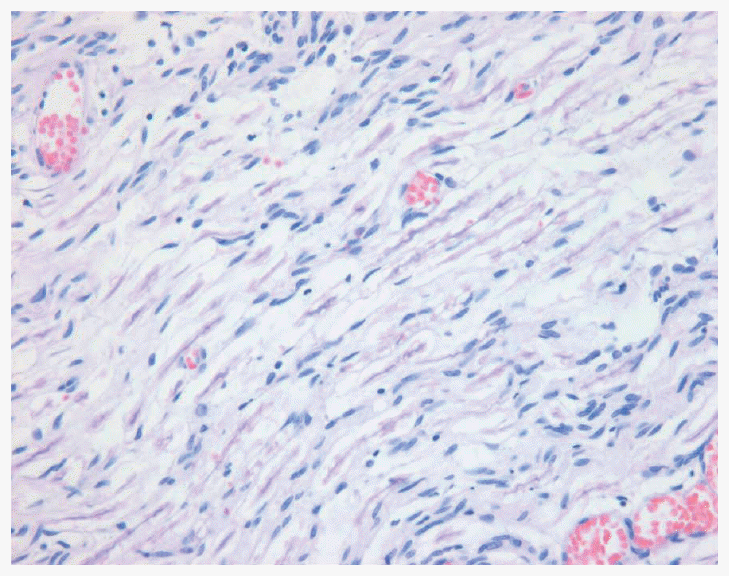
- Further investigations revealed normal plain radiographs with minimal enlargement of the spinal canal in the anteroposterior diameter. In the cervical MRI, we observed a huge lobulated lesion with iso-signal intensity lesion in T1-weighted imaging (T1WI); however, T2-weighted imaging (T2WI) in the ventrolateral aspect revealed heterogeneous high signal intensity lesion, that was pushing the cord to the left dorsal side and extending from C2 to C7 (Figure 1A). The lesion showed no peripheral rim of contrast enhancement. Axial T2WI showed a dumb-bell shaped mass lesion growing through the vertebral foramen (Figure 1B). The intradural extramedullary lesion was approached through a laminectomy of C2 to C7. The mass was found to be attached to redundant nerve roots at both rostral and caudal ends and it grew through the vertebral foramen. The tumor was incompletely removed. Biopsy of the tumor revealed that it was a schwannoma. Histological analysis of the tumor revealed stellate spindle-shaped cells with small, hyperchromatic nuclei, tenuous cytoplasmic processes, and scanty surrounding reticulin were loosely arranged in myxoid stroma. We also observed thickened blood vessels with hyaline deposition on the walls and clumps of hemosiderin-laden foamy macrophages (Figure 2). After the operation, the patient’s tremor improved slightly, but she was prone to falls and unable to walk without a walker.
Case
- Approximately 30% of primary intraspinal tumors originate from Schwann cells. While both schwannomas and neurofibromas are derived from Schwann cells, they show some distinguishing characteristics. They are usually solitary lesions, except in Von Recklinghausen’s disease. Schwannomas are usually solid or heterogeneously solid tumors and are commonly observed as intradural extramedullary spinal tumors.3,4 We have described a case of lobulated Schwannoma in the cervical region that intradurally extended over a long segment from C2 to C7 and also showed extradural extension. Schwannomas are benign tumors with a slow growth rate, the diagnosis of extracranial schwannomas may pose a challenge to neurologists. Patients with such schwannomas rarely show pain and neurological symptoms, and these symptoms become evident only at the advanced stages.4 This case showed a striking paucity of symptoms in spite of the large size of the tumor. Preoperative diagnosis is sometimes difficult and differential diagnoses are widely variable. Surgical treatment does not always fully eliminate the tumor, the possibility of neurological sequelae should be explained to the patient during the preoperative interview.4
- Clonus, presented behaviorally as rhythmic distal joints oscillation, is a common pathology that occurs secondary to spinal cord injury and other neurological disabilities.5 The underlying mechanism of clonus is therefore still unclear and controversial. The prevailing one is that clonus results from recurrent activation of stretch reflexes. Szumski et al. demonstrated that unsustained clonus could be prolonged by Jendrassik’s maneuver.6 They concluded that in clonus the spindles were abnormally sensitive and that the motoneurons particularly important for manifesting clonus were the dynamic fusimotor neurons. Hagbarth et al. demonstrated that Ia afferent discharges precede clonic EMG bursts but were not activated during muscle contraction.7 They concluded that as the muscle relaxes, muscles spindle stretches and regenerates EMG activity resulting in repeated oscillatory movement. Rack et al. also supported the notion that clonus is caused by self-sustaining oscillation of a stretch reflex pathway because the frequency of clonus may be altered by changing the mechanical load acting at the joint.8 An alternative hypothesis is that clonus results from the action of central oscillator. Walsh and Wright have reported similar frequencies of clonus among ankle, knee, and wrist muscles.9 Dimitrijevic et al. reported that ankle clonus frequency was unchanged by altering the frequency of tendon taps, cooling, adding a mass to the foot, moving the foot through viscous solution, and direct movement opposition.2 This study suggested clonus to be primarily dependent on the activity of a central generator within spinal cord which rhythmically activates the alpha motoneurones. The other study showed the mechanism underlying clonus after spinal cord injury is not solely related to recurrent muscle stretch. They concluded that clonus is the frequent presentation of intrinsic oscillating spinal neuronal networks after chronic loss of supraspinal input and diminution of load-related sensory input.5 Therefore, we also suggest that the cervical schwannoma observed in our case could be responsible for deprivation of supraspinal control of the stretch reflex loops.
- The symptoms in this patient were considered similar to action tremors in which rhythmic oscillations of a limb occur spontaneously during voluntary movement. Although action tremors are usually observed in cerebellar, task-specific, dystonic, or Holmes tremor, and they may be rarely associated with partial lesions in the descending motor pathways.10 However, our patient had spasticity in the upper limbs with exaggerated tendon jerks and passive stretch-induced clonus. The case we report is very similar to the description of “action induced clonus mimicking tremor” by Fravix et al.10 However, in our case, duration of the symptom was as long as the essential tremor. This point induced a clinician to insufficient neurologic examination and misdiagnosis. Usually clonus is rarely misinterpreted as tremor. On clinical examination, passive stretching of the muscles increases the force of clonus but not of tremor.12 Even though essential tremor is a common disease, clinician have to do sufficient neurologic examination considering differential diagnosis.
Discussion
Figure 1Cervical MRI. A: Saggital T1-weighted imaging showing a huge lobulating iso-signal intensity intradural extramedullary lesion; T2-weighted imaging in the ventrolateral aspect revealed heterogeneous high signal intensity lesion, that was pushing the cord to the left dorsal side and extending from C2 to C7. B: Axial T2-weighted imaging showing a dumb-bell shaped mass lesion growing through the vertebral foramen.


Figure 2Fascicles of spindle-shaped cells are interrupted by small myxoid areas. Note the small blood vessels with markedly thickened walls.


- 1. Rossi A, Mazzocchio R, Scarpini C. Clonus in man: a rhythmic oscillation maintained by a reflex mechanism. Electroencephalogr Clin Neurophysiol 1990;75:56–63.ArticlePubMed
- 2. Dimitrijevic MR, Nathan PW, Sherwood AM. Clonus: the role of central mechanisms. J Neurol Neurosurg Psychiatry 1980;43:321–332.ArticlePubMedPMC
- 3. Conti P, Pansini G, Mouchaty H, Capuano C, Conti R. Spinal neurinomas: retrospective analysis and long-term outcome of 179 consecutively operated cases and review of the literature. Surg Neurol 2004;61:34–43.ArticlePubMed
- 4. Langner E, Del Negro A, Akashi HK, Araújo PP, Tincani AJ, Martins AS. Schwannomas in the head and neck: retrospectively analysis of 21 patients and review of the literature. Sao Paulo Med J 2007;125:220–222.ArticlePubMedPMC
- 5. Beres-Jones JA, Johnson TD, Harkema SJ. Clonus after human spinal cord injury cannot be attributed solely to recurrent muscle-tendon stretch. Exp Brain Res 2003;149:222–236.ArticlePubMedPDF
- 6. Szumki AJ, Burg D, Struppler A, Velho F. Activity of muscle spindles during muscle twitch and clonus in normal and spastic human subjects. Electroencephalogr Clin Neurophysiol 1974;37:589–597.ArticlePubMed
- 7. Hagbarth KE, Wallin G, Löfstedt L, Aquilonius SM. Muscle spindle activity in alternating tremor of Parkinsonism and in clonus. J Neuro Neurosurg Psychiatry 1975;38:636–641.ArticlePubMedPMC
- 8. Rack PM, Ross HF, Thilmann AF. The ankle stretch reflexes in normal and spastic subjects. The response to sinusoidal movement. Brain 1984;107:637–654.ArticlePubMed
- 9. Walsh EG, Wright GW. Patellar clonus: an autonomous central generator. J Neurol Neurosurg Psychiat 1987;50:1225–1227.ArticlePubMedPMC
- 10. Deuschl G, Raethjen J, Lindemann M, Krack P. The pathophysiology of tremor. Muscle Nerve 2001;24:716–735.ArticlePubMed
- 11. Fraix V, Delalande I, Parrache M, Derambure P, Cassim F. Action-induced clonus mimicking tremor. Mov Disord 2008;28:285–288.Article
- 12. Deuschl G, Bain P, Brin M. Consensus statement of the Movement Disorder Society on Tremor. Ad Hoc Scientific Committee. Mov Disord 1998;13( Suppl 3):2–23.Article
REFERENCES
Figure & Data
References
Citations
Citations to this article as recorded by 

Comments on this article
 KMDS
KMDS
 E-submission
 PubReader
 ePub Link
 Cite
Cite


