Oculogyric Crisis Associated with Disulfiram-Induced Pallidonigral Lesion
Article information
Abstract
We report a man who developed oculogyric crisis one month after disulfiram intoxication. Brain MRI showed lesions involving bilateral globus pallidus and left substantia nigra. In our patient, neuronal discharges from pathologically reorganized basal ganglia circuit to the mid-brain ocular motor center might lead to tonic deviation of the eyes.
Oculogyric crisis (OGC) is characterized by a transient forceful upward conjugate deviation of the eyes. It can be seen in patients with postencephalitic parkinsonism,1 neuropetic induced acute or tardive dystonia,2 Wilson’s disease,3 and lesions involving uni-or bilateral lentiform nuclei.4,5
Disulfiram intoxication may produce a lesion at the globus pallidus and putamen, and cause parkinsonism and dystonia.6,7 We describe a male patient who attempted suicide with a high dose of disulfiram and developed oculogyric crisis, in addition to parkinsonism and dystoina. MRI brain scan studies showed lesions in the bilateral globus pallidus and left substantia nigra.
Case Report
A 15-year old boy attempted suicide by taking a high dose of disulfiram (20 mg). Upon arrival at the hospital, he was alert and vital signs were stable. He had not been treated neuroleptics. One month after, he suddenly developed an acute insult consisted of anxiety attack and subsequent retrocollis and forceful upward deviation of the eyes. During the attack, he could not close his eyes and speak, making only grunting noises. He could not move his eyes voluntarily at all, but horizontal oculocephalic reflexes could be elicited. The attack was occasionally accompanied by dystonic posturing of all four limbs. The frequency, duration and intensity of the attacks increased gradually over a period of eight years, occurring more than 10 times a day and lasted up to 2 hours. Neurological examinations between the attacks revealed mild rigidity of the right arm and leg. There was a delay in initiation of saccadic eye movements. He had no motor weakness or sensory deficits, but he needed a help to walk due to foot dystonia. Deep tendon reflexes were normoactive and symmetric. Plantar reflexes were flexor bilaterally. Routine laboratory tests were all normal. Ictal electroencephalography studies were unremarkable. T2-weighted brain MRI studies showed high signal intensity lesions at the bilateral globus pallidus and left substantia nigra (Figure 1). Levodopa treatment was not effective, but the frequency of the attacks decreased markedly after trihexyphenidyl hydrochloride (2.5 mg tid) treatment. Also, the foot dystonia improved partially and the patient could walk a short distance without assistance.
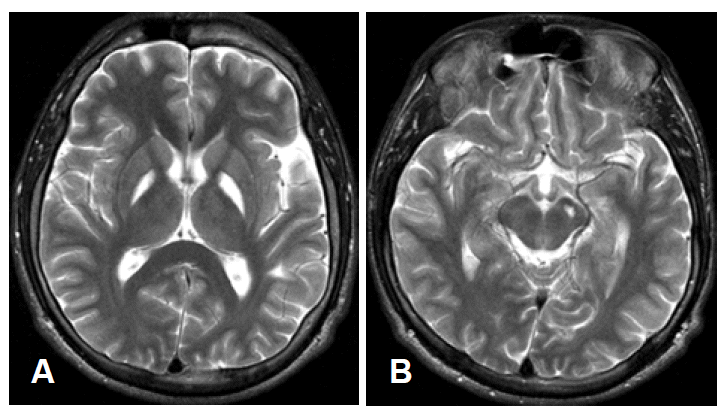
T2-weighted brain MRI shows increased signal intensity in both globus pallidus (A) and left midbrain adjacent substantia nigra (B).
Discussion
Disulfiram is metabolized to cyanide disulfide (CS2) and produce lesions at the globus pallidus and substantia nigra pars reticulata in monkeys.7 In huans, pallidal or lenticular lesions after disulfiram intoxication have been reported.6,7 However, to our knowledge, lesion at the substantia nigra after disulfiram intoxication is rarely documented on brain MRI studies.
OGC occurs frequently in association with neuroleptic treatment, postencephalitic parkinsonism, and focal brain lesions at the putamen or globus pallidus.1,2,4,5 These findings suggest that OGC can be caused by basal ganglia dysfunction, particularly of the dopaminergic system. In our patient, neuronal discharges from pathologically reorganized basal ganglia circuit to the midbrain ocular motor center might lead to tonic deviation of the eyes.
We reported a patient who developed disulfiram induced parkinsonism, dystonia, and OGC, responding markedly to the anticholinergics treatment.