Ventricular Bigeminy after Subcutaneous Administration of Apomorphine in a Patient with Refractory Parkinson’s Disease: A Case Report
Article information
Abstract
Apomorphine is a well established treatment for the management of sudden, unexpected and refractory levodopa-induced “off” states in fluctuating Parkinson’s disease either as bolus injections or as continuous infusions. Incidents of atrial fibrillation associated with the administration of the drug have been reported in the past but no incidents of ventricular arrhythmias. We report a case of ventricular bigeminy recorded in a female patient after the administration of apomorphine.
Parkinson’s disease (PD) is a movement disorder. The most common clinical findings are resting tremor, rigidity and bradykinesia. The evolution is slowly progressive. The treatment that has satisfactory response to a great number of patients with PD is L-Dopa and dopamine receptor antagonists.1
Apomorphine is a high potent D2-, D3- and D4- dopamine receptor agonist with a particularly high D1-dopamine receptor affinity.2 It is used to treat “off” episodes of patients with PD (times of difficulty moving, walking and speaking that may happen as medication wear off). Due to its almost complete inactivation during liver passage it is applied subcutaneously. Its effect is waning after about an hour.3,4 The most common side effects are cutaneous nodules at the injections sites, nausea, headache, diarrhoea, weakness and orthostatic hypotension.5 Hallucinations, chest pain and visual disturbances may occur. The daily dose ranges from 3 to 30 mg.6
We present a case of a relatively rare benign cardiac arrhythmia that occurred after the administration of apomorphine to a senior female patient.
Case
An 87 years old woman with a history of PD for the last 18 years, treated with L-Dopa and dopamine receptor antagonists, presented with “off” episodes during the last two months despite combined medical therapy. At presentation she was treated with carbidopa 87.5 mg/day, levodopa 875 mg/day, pramipexol 1.4 mg/day and mitrazapine 7.5 mg/day. She was administered to the Neurology Department for scheduled initiation of apomorphine treatment.
There was no other medical history recorded, despite the advanced age of the patient, and the standard electrocardiogram (ECG) (Figure 1) at her admission was at normal range. Full blood tests, including liver and renal function and electrolytes’ levels, were conducted and no abnormal values were detected.

The first electrocardiogram shows normal findings.
The previous medical therapy was discontinued 48 hours before the initiation of apomorphine, according to the usual practice.
The patient was haemodynamically stable before the initiation of apomorphine and the heart rate and blood pressure was repeatedly monitored.
Ten minutes after the first subcutaneous administration of 10 mg of apomorphine, a heart rhythm disturbance was observed and recorded in a 12-lead ECG. The disturbance was ventricular bigeminy (Figure 2), with a left bundle branch block morphology of the ventricular origin QRS complexes and a frontal QRS axis at −10°. No haemodynamic instability was recorded and no symptom (dizziness, palpitations, pre-syncope or syncope) was reported by the patient. The patients’ heart rhythm and blood pressure was monitored repeatedly during the following hours.
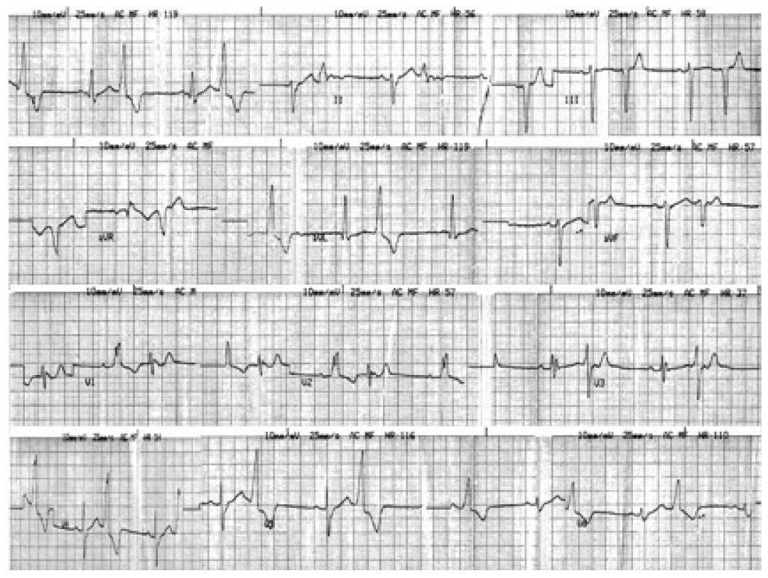
The electrocardiogram just after the first injection of apomorphine shows ventricular bigeminy with left bundle brunch block.
The next day the 12-lead ECG showed ventricular bigeminy again, so we decided to monitor the patients’ heart rhythm with a 24-hour ambulatory recorder (Holter). The recording showed brief periods of ventricular bigeminy (Figure 3) and no other arrhythmias. We conducted a two dimensional echo-cardiogram without any pathological findings that could pre-dispose to the emergence of a ventricular arrhythmia (e.g., ventricular hypertrophy or any indication of ischemic heart disease), and three days later we repeated the 24-hour ambulatory recording, which revealed no heart rhythm disturbances at all.

The 24-hour ambulatory recorder one day after the apomorphine injection shows ventricular bigeminy.
The patient returned to the previous treatment for PD as it is mentioned above, and no such arrhythmia was recorded in the follow up visits of the patient.
Discussion
This was a rare case of heart rhythm disturbance associated with apomorphine treatment for PD.7 We conducted a review of related literature in PubMed and Medline and no such case was ever reported. The available drug information does not mention such an arrhythmia as a possible adverse event related to the drug administration. The QT interval prolongation is the only electrocardiographic disturbance previously referred,8 but in our patient no change in QT interval was observed.
The aetiology of ventricular bigeminy in this setting is difficult to determine. The advanced age of the patient might be a predisposing factor to cardiac arrhythmias,9 although there was no previous history or symptoms of cardiac disease reported nor any structural abnormality detected.
Apomorphine is a potent dopamine receptor agonist, and it is known that other agonists like dopamine may increase the Purkinje fibers automaticity and have a biphasic effect on action-potential duration causing atrial and ventricular tachyarrhythmias.10 Yet, there is no evidence that such a mechanism is responsible for the reported incident of bigeminy. Besides, the affinity of apomorphine to the β1 adrenergic receptors through which dopamine exerts its cardiac effects is very low.11
Although the arrhythmia was self terminated and didn’t cause any symptoms or haemodynamic instability, it would be reasonable to monitor inpatients closely for the emergence of cardiac dysrythmias at the initiation of the treatment with apomorphine and during dose titration. As far as outpatients are concerned, regular follow up for the emergence of cardiac arrhythmias should be recommended.