Parkinsonism and Dementia Associated with Giant Virchow-Robin Spaces
Article information
Virchow-Robbin space (VRS) denotes dilated subarachnoid space along the penetrating arteries to the level of capillaries. Giant VRSs (GVRS), defined as greater than 1.5 cm, are found in the basal ganglia along the lenticulostriate arteries, pons and midbrain along the collicular arteries and high cerebral convexity along the medullary arteries [1,2].
CASE HISTORY
Most patients with GVRSs in the high cerebral convexity exhibit no neurological deficits [1,3-5], including motor and sensory evoked potential studies [3]. We present a patient who developed parkinsonism and dementia associated with GVRSs. Positron emission tomography (PET) studies showed normal striatal dopamine transporter uptake, but reduced glucose metabolism in the cerebral cortex and right thalamus.
A 64-year-old man developed bradykinesia and memory disturbances. On neurological examination, the patient was found to have a masked face. Glabellar and snouting reflexes were present. Speed and amplitude of finger and foot tapping were reduced bilaterally and were pronounced on the left side. Muscle tone was mildly increased in all four limbs tested. On pull tests, the patient stabilized after 3 to 4 backwards steps. He stood on widened base and walked with mildly reduced stride and cadence. The patient’s Unified Parkinson’s Disease Rating Scale (UPDRS) total motor score was 15. There were no abnormalities on cerebellar function tests. Mini-Mental State Examination (MMSE) score was 27. Neuropsychological tests, however, showed impairments (< 15 percentile for age and sex matched controls) in immediate and delayed recall of verbal and visual subjects, attention, confrontational naming, generative naming, visuospatial function, and inhibitory control. T2 weighted brain magnetic resonance imaging (MRI) studies showed scattered high signal intensity and multiple round and septate cystic lesions, mainly in the right parietal, frontal and temporal white matters (Figure A, B, and C). MR cerebral angiography showed no abnormalities. [18F]-FP-CIT PET studies showed normal striatal uptake. [18F]-deoxyglucose PET studies showed hypometabolism, predominantly involving the right thalamus and the right parietal, frontal and temporal cortical areas (Figure D, E, and F). On follow-up examination 4 years after onset, there was worsening of parkinsonian motor deficits, particularly gait disturbances and postural instability, and further cognitive dysfunctions. His UPDRS total motor score was 27 and MMSE score was 20. Follow-up brain MRI studies showed no significant changes.
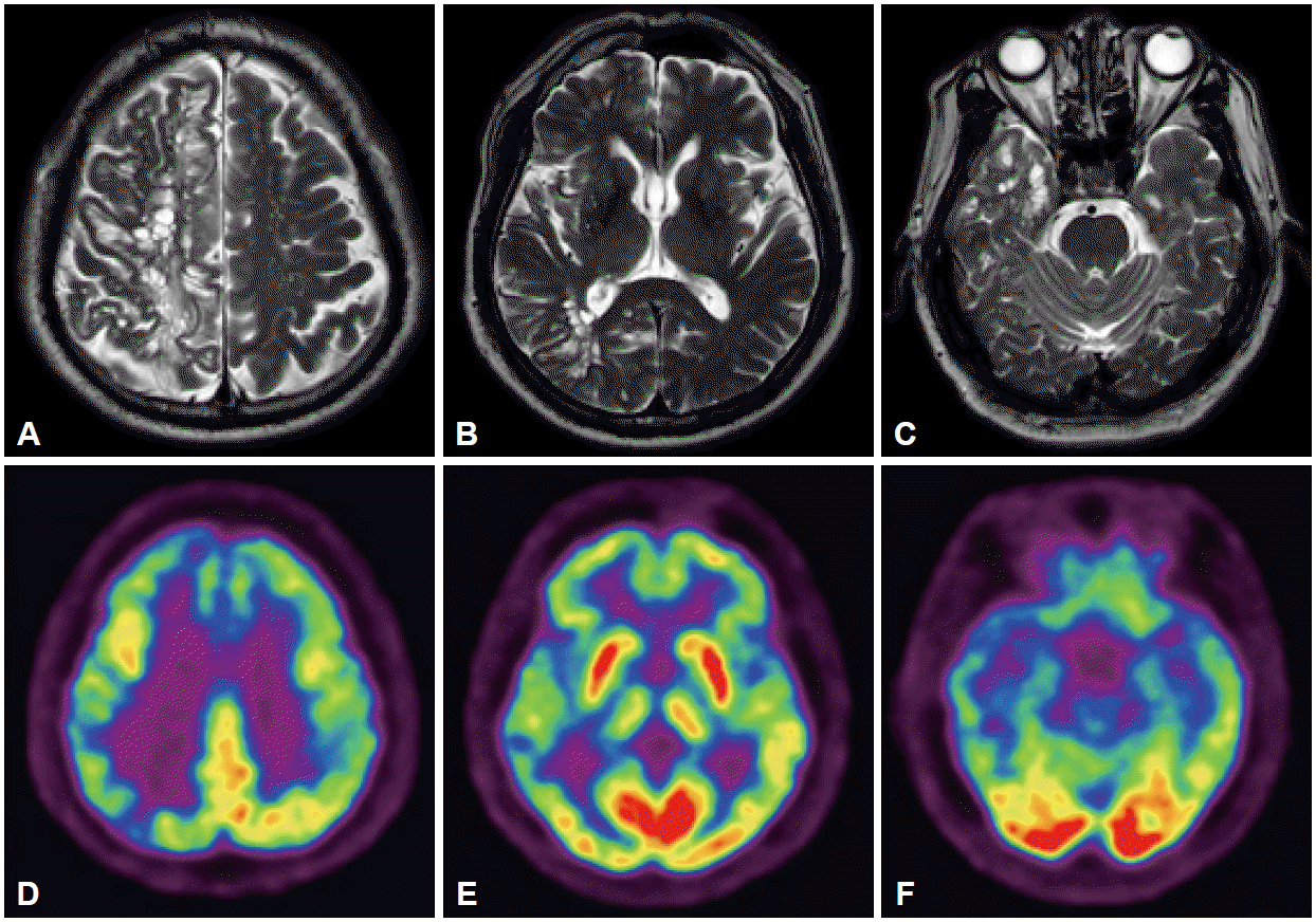
T2-weighted brain magnetic resonance imaging studies show mixture of high signal intensity lesions and numerous round and septate cystic lesions in the (A) right high cerebral convexity and (B and C) temporal lobes. [18F]-deoxyglucose PET studies show diffuse cerebral cortical hypometabolism, predominantly in the (D) bilateral frontal, right parietal (E) right thalamic and (F) right temporal cortices. PET: positron emission tomography.
DISCUSSION
Giant Virchow-Robbin spaces in the high cerebral convexity are characterized by clustered sharp demarcated round, oval, or linear cystic lesions. On brain MRI studies, they did not enhance and are iso-intense with cerebrospinal fluid [2,3]. In approximately 80% of reported cases, GVRSs involved one cerebral hemisphere [1-5].
Giant Virchow-Robbin spaces in the high cerebral convexity are usually found incidentally [4,5] or during the evaluation for non-specific neurological symptoms (e.g., fainting, dizziness, hearing impairment [1], headache, or visual change [2]). Rarely, patients may present with dementia [2]. The patient reported herein developed progressive parkinsonism and dementia in association with GVRSs in the high cerebral convexity.
MR tractography studies of patients with GVRSs showed markedly reduced fibers in the affected cerebral hemisphere [4] Therefore, GVRSs and white matter lesions in our patient may have disrupted cortical efferent fibers to the basal ganglia and afferent fibers to the motor-related cerebral cortices. The parkinsonism seen in our patient seemed to share a common pathogenic mechanism with the parkinsonism associated with Binswanger disease. Alternatively, right thalamic hypometabolism, representing reduced basal ganglia or cortical inputs to the thalamus, may also have contributed to the asymmetric parkinsonism of our patient.
In our patient, cortical glucose hypometabolism and consequent cognitive dysfunctions seemed to be associated with loss of afferent fibers to the cerebral cortex or cortical neuronal loss associated with retrograde axonal degeneration. Our patient demonstrated that GVRSs in the high cerebral convexity are not always clinically silent but may cause progressive parkinsonism and dementia.
Notes
Conflicts of Interest
The authors have no financial conflicts of interest.