Giant Middle Fossa Epidermoid Presenting as Holmes’ Tremor Syndrome
Article information
Abstract
Intracranial dermoids may gradually reach an enormous size before the onset of symptoms. Common clinical presentations of intracranial epidermoid include headache and seizures. We present a case of a 35-year female patient with giant middle fossa epidermoid that presented with Holmes’ tremor syndrome, and we review the relevant literature. To the best of our knowledge, such a presentation has not previously been described in the literature.
Intracranial dermoids may gradually reach an enormous size before the onset of symptoms.1,2 Common clinical presentations of intracranial epidermoid include headache and seizures,3,4 and the latter can be important clinical presenting features in patients in which the tumor is in proximity to the temporal lobe.5 We present a case of a female patient with a giant middle fossa epidermoid that presented with Holmes’ tremor syndrome; we also review the relevant literature.
CASE
A right-handed, 35-year-old female presented with headache and involuntary movements involving her right upper and lower limbs of two-month duration. Her general physical and systemic examination was unremarkable. On examination, the patient had a coarse tremor that was slow, at a frequency of approximately 2–4 Hz. When the patient held her hands out in front of her, the amplitude grossly increased, with some amount of distal posturing. The tremor also became prominent proximally. During attempts at holding objects or reaching for targets, the tremors were more prominent, with irregular arrhythmic and jerky components. The tremors were more prominent in the arm. Her higher mental functions, cranial nerve, sensory, and deep tendon reflex examinations were normal. Cerebellar examination was difficult because of the tremors; however, there was no nystagmus, and speech and gait were normal. Chest X-ray and electrocardiogram were normal. Contrast enhanced CT scans of her brain revealed a large non-enhancing well-marginated extra-axial mass in the left fronto-tempo-parietal region. There was a significant mass effect and midline shift (Figure 1). Based on the imaging findings, a large epidermoid cyst presenting with movement disorder was suspected. The patient underwent left fronto-temporo-parietal craniotomy and decompression of the tumor. During surgery, there was a large avascular mass with pearly white contents. The mass could be completely removed. Histopathological examination of the mass revealed keratinizing squamous epithelium with keratinous debris arranged in laminated layers. Follow-up CT scan showed complete removal of the tumor (Figure 2). At 1 year follow-up, the patient had improved significantly: her ataxia was completely relived, and there was no headache or vomiting.

Pre-operative plain CT scan brain showing non-enhancing hypodense extensive mass lesion in the left fronto-temporo-parietal area with mass effect and midline shift.
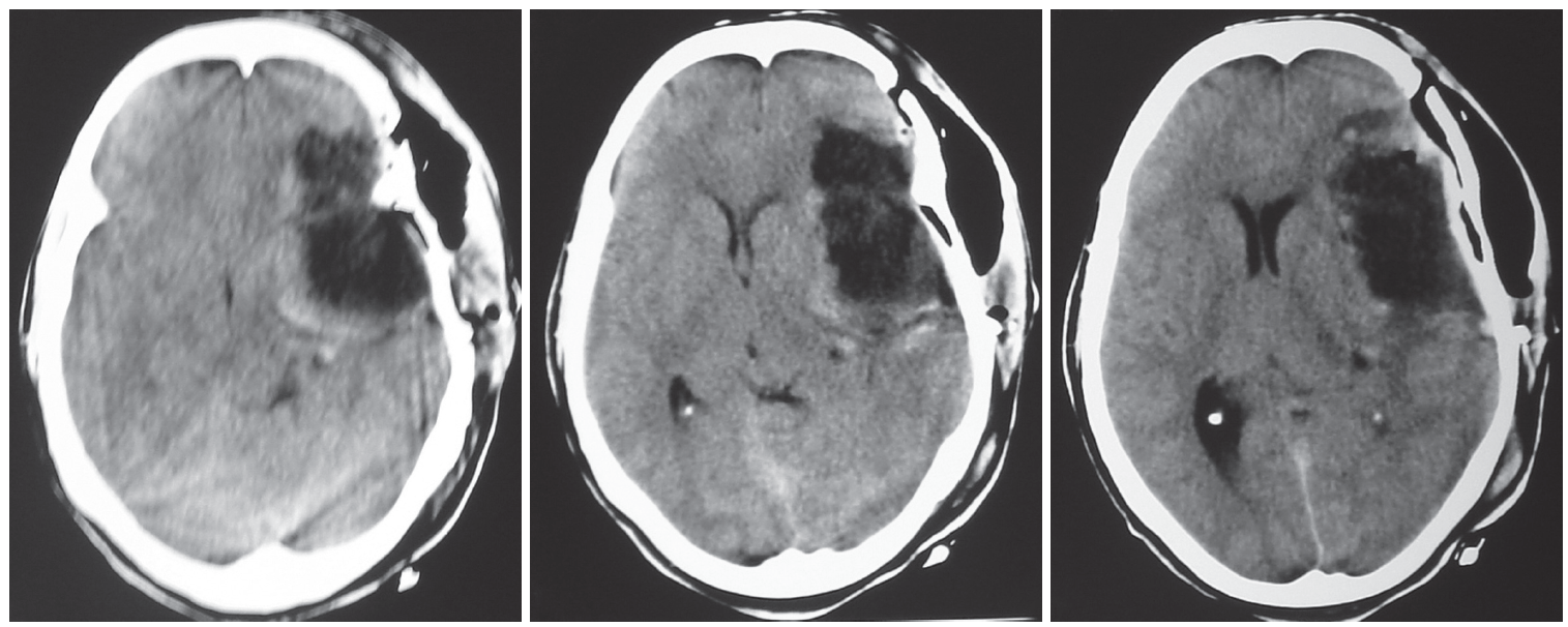
Follow-up scan showing reduction in the mass effect and midline shift.
DISCUSSION
Intracranial space-occupying lesions in different locations, including tumors either because of mass effect causing distortion of the connecting pathways and vascular compromise or direct infiltration, have been increasingly recognized as a cause of a spectrum of movement disorders.6–8 Holmes tremor (previously known as rubral or midbrain tremor) has been recognized as a distinct clinical entity9,10 and was described by Gorden Holmes.11 Holmes, tremor syndrome is characterized by an irregular low-frequency rest and intention tremor, and in many cases, there is also a postural tremor.9 These tremors can be exacerbated by postural adjustments and by guided voluntary movements.12 Lesions of the superior cerebellar peduncle, midbrain tegmentum or posterior part of the thalamus may cause this peculiar tremor, and it is probable that lesions of the red nucleus itself are not crucial for its production.12 It has been postulated that Holmes, tremor syndrome arises from lesions that interrupt the dentate-thalamic and the nigrostriatal tracts, thus causing both an action and a rest tremor.9,13 These tremors can be generated in lesions of the cerebello-rubro-thalamic system without evidence of a rubral lesion itself.14 In our case, the tremors were also present in rest, posture and intention, with distal and proximal components, and best fit the diagnosis of rubral tremor. In our case, there was most likely extrinsic compression of the midbrain tegmentum, as observed in Figure 1. As it was an extrinsic lesion, the decompression helped for partial relief of the tremor. In summary, intracranial epidermoids are slow-growing benign, congenital, developmental tumors that tend to occur in the cerebellopontine angle, cerebellar vermis, fourth ventricle, parasellar region and frontal and fronto-temporal cisterns.15 Although intracranial epidermoids can be suspected on brain imaging, i.e., both CT and MRI, fast fluid-attenuated inversion recovery16 and diffusion-weighted MR imaging is more sensitive than conventional MR imaging.17,18 Whenever possible, complete excision of the epidermoid is the treatment of choice.19 As in the present case, the complete removal of the tumor will not only remove the tumor tissue but also can reverse the clinical findings (complete abolition and/or better control of tremors).7
Notes
Conflicts of Interest
The authors have no financial conflicts of interest.