Skip Navigation
Skip to contents

JMD : Journal of Movement Disorders

OPEN ACCESS
SEARCH
Search

Articles

Page Path
HOME > J Mov Disord > Volume 10(1); 2017 > Article
Letter to the editor
Camptocormia with Transient Ischemic Attack
Ju-Hee Oh, Dong-Woo Ryu, Si-Hoon Lee, Joong-Seok Kim
Journal of Movement Disorders 2017;10(1):62-63.
DOI: https://doi.org/10.14802/jmd.16043
Published online: January 18, 2017

Department of Neurology, College of Medicine, The Catholic University of Korea, Seoul, Korea

Corresponding author: Joong-Seok Kim, MD, PhD, Department of Neurology, Seoul St. Mary’s Hospital, College of Medicine, The Catholic University of Korea, 222 Banpo-daero, Seocho-gu, Seoul 06591, Korea / Tel: +82-2-2258-6078 / Fax: +82-2-599-9686 / E-mail: neuronet@catholic.ac.kr
• Received: September 19, 2016   • Revised: October 17, 2016   • Accepted: October 31, 2016

Copyright © 2017 The Korean Movement Disorder Society

This is an Open Access article distributed under the terms of the Creative Commons Attribution Non-Commercial License (http://creativecommons.org/licenses/by-nc/3.0/) which permits unrestricted noncommercial use, distribution, and reproduction in any medium, provided the original work is properly cited.

  • 7,097 Views
  • 83 Download
  • 1 Web of Science
  • 2 Crossref
Dear Editor,
Camptocormia is a condition characterized by an abnormal flexion of the thoracolumbar spine, which appears while standing or walking. It is associated with several clinical conditions, such as parkinsonism, dystonia and neuromuscular disorders [1]. In addition, camptocormia is a side effect of neuroleptic and anti-parkinsonian drugs. In rare cases, patients with trauma, arthritis, or malignancy may present camptocormia [2].
We present the case of a 66-year-old female who had transient and repeated camptocormia with an acute onset. Five days prior to presentation, episodes of marked camptocormia during standing and walking began. The patient reported that she was unable to maintain upright posture (nearly 80° painless forward flexion) and her gait festinated. Her symptoms spontaneously disappeared without any medical treatment after approximately twenty minutes but reoccurred three more times. The patient had hypertension and diabetes mellitus that were not well controlled. She did not have a history of smoking. She had not taken any antipsychotic medications other than an angiotensin II receptor blocker.
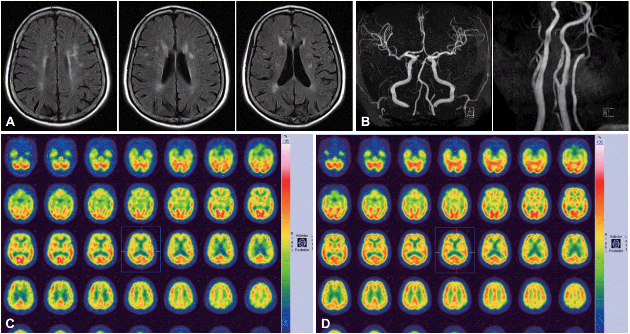
Upon admission, no abnormalities were found on her physical and neurological examination. The results from laboratory tests for complete blood cell and platelet count, erythrocyte sedimentation rate, blood electrolytes, creatinine, liver enzymes, cholesterol, triglycerides, creatinine phosphokinase, lactate dehydrogenase, prothrombin, and partial thromboplastin time were normal. In addition, autoantibody screens for vasculitis were also normal. Magnetic resonance imaging of her brain revealed multiple hyperintensities in both subcortical white matter, while magnetic resonance angiography showed no definite intracranial and extracranial stenoses (Figure 1). Neither an electrocardiogram nor a transthoracic echocardiogram revealed cardiac abnormalities. A 99mTc-ethylcysteinate dimer single photon emission computed tomography (SPECT) scan of the brain both at baseline and after acetazolamide showed a decrease in cerebral blood flow in the left parietal cortex without vascular reserve (Figure 1). No abnormalities were found using electromyography on the cervical and thoracolumbar muscles.
After the patient was treated with 100 mg aspirin, transient camptocormia disappeared during a 10-month follow-up period.
Although the pathophysiology of camptocormia is not currently clear, two provisional pathomechanisms have been suggested. Camptocormia could result from central neurodegenerative disorders, principally Parkinson’s disease, or peripheral mechanisms associated with primary musculo-skeletal disorders [1-3].
There have been few reports of secondary and acute camptocormia associated with putaminal vascular lesions [4]. In this case study, the patient complained of transient and repeated camptocormia. We cannot be entirely sure of other conditions for the transient occurrence of the patient’s symptoms, such as epileptic or psychological origins; however, we believe that vascular etiology may be fit to explain this phenomenon from a nosological perspective. The mechanism underlying camptocormia in our patient is unknown. Although ischemic lesions were not found, SPECT imaging of her brain revealed decreased perfusions in the left parietal cortex. Somatosensory, vestibular, and visual sensations are integrated at the temporoparietal and posterior parietal cortices. The posterior parietal cortex and its connection to the frontal lobe play an essential role in maintaining body schema [5,6]. Thus, potential explanations for camptocormia in this patient are transient ischemia in the left parietal lobe and impairment in the parietofrontal connection for anticipatory postural control [7].
In summary, this case suggests that vascular disturbances of the parietal cortex may cause camptocormia. Taken together, this case may aid in our understanding of the relationship between brain lesions and the etiology of camptocormia, although a cause-and-effect relationship cannot be completely proven.

Conflicts of Interest

The authors have no financial conflicts of interest.

Figure 1.
Magnetic resonance imaging of the patient’s brain showed multiple hyperintensities in subcortical white matter (A) and magnetic resonance angiography showed no definite intracranial and extracranial stenoses (B). 99mTc-ethylcysteinate dimer single photon emission computed tomography scan of the brain at baseline (C) and after acetazolamide (D) showed a decrease in cerebral blood flow in the left parietal cortex without vascular reserve.
jmd-16043f1.gif
  • 1. Lenoir T, Guedj N, Boulu P, Guigui P, Benoist M. Camptocormia: the bent spine syndrome, an update. Eur Spine J 2010;19:1229–1237.ArticlePubMedPMC
  • 2. Finsterer J, Strobl W. Presentation, etiology, diagnosis, and management of camptocormia. Eur Neurol 2010;64:1–8.ArticlePubMed
  • 3. Srivanitchapoom P, Hallett M. Camptocormia in Parkinson’s disease: definition, epidemiology, pathogenesis and treatment modalities. J Neurol Neurosurg Psychiatry 2016;87:75–85.ArticlePubMed
  • 4. Nieves AV, Miyasaki JM, Lang AE. Acute onset dystonic camptocormia caused by lenticular lesions. Mov Disord 2001;16:177–180.ArticlePubMed
  • 5. Takakusaki K. Neurophysiology of gait: from the spinal cord to the frontal lobe. Mov Disord 2013;28:1483–1491.ArticlePubMed
  • 6. Lajoie K, Drew T. Lesions of area 5 of the posterior parietal cortex in the cat produce errors in the accuracy of paw placement during visually guided locomotion. J Neurophysiol 2007;97:2339–2354.ArticlePubMed
  • 7. Park JH, Kang YJ, Horak FB. What is wrong with balance in Parkinson’s disease? J Mov Disord 2015;8:109–114.ArticlePubMedPMCPDF

Figure & Data

References

    Citations

    Citations to this article as recorded by  
    • Mesial Frontal Lobe Infarction Presenting as Pisa Syndrome
      Kazuyuki Noda, Maya Ando, Takayuki Jo, Anri Hattori, Kotaro Ogaki, Mizuho Sugiyama, Nobutaka Hattori, Yasuyuki Okuma
      Journal of Stroke and Cerebrovascular Diseases.2020; 29(8): 104882.     CrossRef
    • Transient camptocormia with citalopram treatment in a patient with mixed dementia–A case report
      Segal Inbal, RN Galia Fisher, Merims Doron
      Archive of Gerontology and Geriatrics Research.2020; : 040.     CrossRef

    Comments on this article

    Add a comment
    Figure

    JMD : Journal of Movement Disorders