 E-submission
Articles
- Page Path
- HOME > J Mov Disord > Volume 9(1); 2016 > Article
-
Original Article
N30 Somatosensory Evoked Potential Is Negatively Correlated with Motor Function in Parkinson’s Disease - Suk Yun Kang1, Hyeo-Il Ma2
-
Journal of Movement Disorders 2016;9(1):35-39.
DOI: https://doi.org/10.14802/jmd.15038
Published online: January 25, 2016
1Department of Neurology, Dongtan Sacred Heart Hospital, Hallym University College of Medicine, Hwaseong, Korea
2Department of Neurology, Hallym University Sacred Heart Hospital, Hallym University College of Medicine, Anyang, Korea
- Corresponding author: Suk Yun Kang, MD, PhD, Department of Neurology, Dongtan Sacred Heart Hospital, Hallym University College of Medicine, 7 Keunjaebong-gil, Hwaseong 18450, Korea / Tel: +82-31-8086-2310 / Fax: +82-31-8086-2317 / E-mail: sukyunkang@hanmail.net
Copyright © 2016 The Korean Movement Disorder Society
This is an Open Access article distributed under the terms of the Creative Commons Attribution Non-Commercial License (http://creativecommons.org/licenses/by-nc/3.0) which permits unrestricted non-commercial use, distribution, and reproduction in any medium, provided the original work is properly cited.
ABSTRACT
-
Objective
- The aim of this study was to investigate frontal N30 status in Parkinson’s disease (PD) and to examine the correlation between the amplitude of frontal N30 and the severity of motor deficits.
-
Methods
- The frontal N30 was compared between 17 PD patients and 18 healthy volunteers. Correlations between the amplitude of frontal N30 and the Unified Parkinson’s Disease Rating Scale (UPDRS) motor score of the more severely affected side was examined.
-
Results
- The mean latency of the N30 was not significantly different between patients and healthy volunteers (p = 0.981), but the mean amplitude was lower in PD patients (p < 0.025). There was a significant negative correlation between the amplitude of N30 and the UPDRS motor score (r = -0.715, p = 0.013).
-
Conclusions
- The frontal N30 status indicates the motor severity of PD. It can be a useful biomarker reflecting dopaminergic deficits and an objective measurement for monitoring the clinical severity of PD.
- Participants
- We recruited PD patients and healthy volunteers who visited the Department of Neurology at Hallym University Sacred Heart Hospital. PD was diagnosed according to the UK Brain Bank criteria [14]. PD patients had either asymmetric or unilateral motor symptoms. To investigate the correlation between motor deficit severity and N30 responses on the more-severely affected side, the UPDRS motor score of the more severely affected side was collected. We selected items 20–26 for the UPDRS, as these items can be measured bilaterally. The scores related to the face in item 20 and the neck in item 22 were not included, as we considered these symptoms axial. We used the UPDRS motor score for only the more severely affected side because if the frontal N30 is associated with motor function, then there should be a clear correlation between frontal N30 and the UPDRS motor score of the more severely affected side. All participants were enrolled in the study after they provided informed consent, and the study protocol was approved by the internal review of Hallym University Sacred Heart Hospital.
- SEP recordings
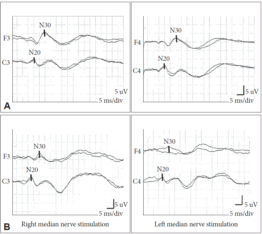
- Median nerve SEPs were recorded from all participants with an EMG machine (Neuropack 8, Nihon-Kohden, Tokyo, Japan). During the recording, participants lay in a quiet, semi-darkened room. Five of the 17 patients were de novo PD (Table 1), and the others were taking their usual medication. Each median nerve was stimulated at the wrist. The stimuli were square-wave electrical pulses with a 0.2-ms duration and applied at a frequency of 2 Hz. The stimulus intensity was determined so that it produced a painless contraction of the abductor pollicis brevis muscle. The amplifier bandpass was 20–3000 Hz.
- SEPs were recorded with Ag/AgCl disc electrodes placed on the scalp at F3/F4, C3/C4 according to the international 10–20 system. Each electrode was referenced to linked earlobes. At least 500 stimuli were applied for each recording. Recordings were performed at least twice to confirm behavior. N20 and P25 waves were recorded from the C3/C4 electrodes, whereas the P22 and N30 waves were recorded from the F3/F4 electrodes. The peak latency was measured from the onset of the stimulus artifact. The peak-to-peak amplitude was also measured.
- Statistical analysis
- Data were expressed as the mean ± standard deviation. The mean age, mean peak latency values, and peak-to-peak amplitudes were compared between PD patients and healthy volunteers with unpaired t-tests. Sex was compared between patients and healthy volunteers with Fisher’s exact test. Spearman’s correlation was used to evaluate the response to stimulation for the more severely affected side and the UPDRS motor score. Values of p < 0.05 were considered significant.
MATERIALS & METHODS
- Seventeen PD patients (mean age: 65.7 ± 8.8 years; 9 women) and 18 healthy volunteers (mean age: 61.1 ± 12.0 years; 11 women) participated in this study. There was no significant difference in age (p = 0.217) or sex (p = 0.738) between the two groups. The clinical features of the PD patients are summarized in Table 1. The UPDRS motor score was obtained from 11 patients.
- The mean latency and amplitude values for the frontal N30 and parietal N20 are shown in Table 2. We obtained the N20 in all patients and controls, but were unable to obtain the N30 on the more-severely affected side in three PD patients. There was no significant difference in the mean latency of N30 between patients and controls (p = 0.981), but the mean amplitude of N30 was lower in PD patients (p = 0.025). No significant differences between patients and controls were observed in the N20 latency and amplitude (Figure 1).
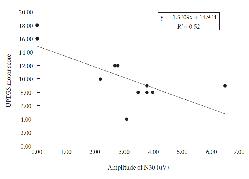
- A correlation analysis was performed in 11 PD patients. The amplitudes of N30 and UPDRS motor scores were significantly and negatively correlated on the more severely affected side (r = -0.715, p = 0.013) (Figure 2).
RESULTS
- Our study demonstrates that the frontal N30 represents the motor dysfunction of PD patients. The amplitudes were lower in PD patients than in healthy volunteers and were closely correlated with motor deficit severity on the more-severely affected side of PD patients.
- The frontal N30 amplitude reflects the functional connectivity of sensorimotor integration, which includes the thalamus, premotor area, basal ganglia and primary motor cortex [15]. The precise location of this activity is still unknown, but it is thought to originate in the SMA [16,17]. Frontal N30 activity was attenuated in meningioma compressing the SMA [18], and intracortical recordings showed that N30 activity was most evident within the premotor cortex [16]. The increase in N30 amplitude seems to result from increased neuronal activity in these regions, as the N30 amplitude increases following repetitive motor tasks [15]. The increase in N30 amplitude may be a result of cortical inhibition rather than facilitation [19].
- Our results (low N30 amplitude in PD) might be due to decreased activity in the SMA. In PD, the activity in the SMA is reduced, but this can be reversed with antiparkinsonian medication (i.e., apomorphine, levodopa) [20,21]. The negative correlation we found between the N30 amplitude and motor deficit severity suggests that the N30 might be associated with dopaminergic activity. In previous studies, the N30 amplitude increased with dopaminergic medications, which also supports our assumption [22,23]. Two such studies indicated the N30 amplitude increased after apomorphine [22] and was positively correlated with plasma levels of levodopa [23].
- As described previously, explanations for the negative results in prior studies of the relationship between PD and motor deficit severity remains unclear. One potential explanation for the negative results is the use of small sample sizes. We addressed this issue in our present study by using larger experimental and control populations than those used in previous investigations. There may be additional explanations for these negative results. Generally, the patient population might be a contributing factor (i.e., age, disease duration, etc.). We summarized the characteristics of the patient populations in previous studies (Table 3). We also speculate that other factors might modulate the amplitude of the frontal N30. The frontal N30 is reduced during motor preparation and planning, observation, and imaginary movement, as well as with sensory inputs including touch and proprioception [24-27]. In fact, because we did not give specific instructions about these factors (i.e., observation, thought) to our patients during SEP recording, we do not know whether these are indeed potential confounders, which remains to be investigated.
- The frontal N30 amplitude presents sensorimotor integration, possibly relating to dopaminergic function. Our study indicates that the frontal N30 amplitude is lower in PD and is negatively correlated with motor deficit severity. The frontal N30 can be a useful ancillary biomarker for monitoring the clinical severity of PD. Additional work is needed to further examine the characteristics of the frontal N30 and its contributing factors.
DISCUSSION
| Parameter | Value |
|---|---|
| Drug-naïve, n | 5 |
| More-severely affected side | |
| Right, n* | 12 |
| Left, n | 5 |
| Disease duration, months (mean ± SD)† | 49.3 ± 40.2 |
| Hoehn & Yahr stage (mean ± SD)‡ | 2.0 ± 0.7 |
| UPDRS motor score (mean ± SD)§ | 10.4 ± 3.8 |
The face of item 20 and the neck of item 22 were not considered. The mean value of UPDRS motor score was averaged from 11 patients.
* right and left designates the more-severely affected side,
† disease duration is mean value of 14 patients,
‡ Hoehn & Yahr stage is mean value in 13 patients,
§ UPDRS motor score of individual patient was the sum of UPDRS items 20‒26 on the more-severely affected side.
UPDRS: Unified Parkinson’s Disease Rating Scale, SD: standard deviation.
| Study | Participants (number) | Age (mean ± SD, year) | Disease duration (mean ± SD, months) | Hoehn & Yahr stage (mean ± SD) | Medication status |
|---|---|---|---|---|---|
| Rossini et al.[9] | PD (14) | 64.6 ± 8.76 | 33.8* | - | On |
| Vascular parkinsonism (2) | |||||
| Healthy (12) | 64.1 ± 9.51 | - | - | - | |
| Garcia et al.[11] | PD (10) | 58.2 ± 12.5 | - | - | On (one patient, de novo) |
| Healthy (10) | 53.4 ± 19.9 | - | - | - | |
| Drory et al.[10] | PD (14) | 63* | 72* | - | Off |
| Healthy (10) | 62* | - | - | - | |
| Bostantjopoulou et al.[12] | PD (23) | 61.6 ± 6.3 | 128.4 ± 55.2 | 2.67 ± 0.4 | On |
| Healthy (23) | 59.8 ± 7.8 | - | - | - | |
| Bostantjopoulou et al.[28] | PD (20) | 65.6 ± 3.8 | 117.6 ± 63.6 | 3 & 4 | On |
| Gironell et al.[5] | PD (8) | 66.25 ± 5.42 | 213 ± 32.4 | - | Off |
| Insola et al.[29] | PD (6) | 53.7 ± 7.5 | - | - | - |
- 1. Daube JR, Rubin DI. Clinical neurophysiology. 3rd ed. New York: Oxford University Press; 2009.
- 2. Thirumala P, Lai D, Engh J, Habeych M, Crammond D, Balzer J. Predictive value of somatosensory evoked potential monitoring during resection of intraparenchymal and intraventricular tumors using an endoscopic port. J Clin Neurol 2013;9:244–251.ArticlePubMedPMC
- 3. Restuccia D, Valeriani M, Barba C, Le Pera D, Bentivoglio A, Albanese A, et al. Abnormal gating of somatosensory inputs in essential tremor. Clin Neurophysiol 2003;114:120–129.ArticlePubMed
- 4. Restuccia D, Valeriani M, Barba C, Le Pera D, Capecci M, Filippini V, et al. Functional changes of the primary somatosensory cortex in patients with unilateral cerebellar lesions. Brain 2001;124(Pt 4):757–768.ArticlePubMedPDF
- 5. Gironell A, Rodríguez-Fornells A, Kulisevsky J, Pascual B, Barbanoj M, Otermin P. Motor circuitry re-organization after pallidotomy in Parkinson disease: a neurophysiological study of the bereitschaftspotential, contingent negative variation, and N30. J Clin Neurophysiol 2002;19:553–561.ArticlePubMed
- 6. Nakata H, Sakamoto K, Yumoto M, Kakigi R. The relationship in gating effects between short-latency and long-latency somatosensory-evoked potentials. Neuroreport 2011;22:1000–1004.ArticlePubMed
- 7. Desmedt JE, Bourguet M. Color imaging of parietal and frontal somatosensory potential fields evoked by stimulation of median or posterior tibial nerve in man. Electroencephalogr Clin Neurophysiol 1985;62:1–17.ArticlePubMed
- 8. Cheron G, Piette T, Thiriaux A, Jacquy J, Godaux E. Somatosensory evoked potentials at rest and during movement in Parkinson’s disease: evidence for a specific apomorphine effect on the frontal N30 wave. Electroencephalogr Clin Neurophysiol 1994;92:491–501.ArticlePubMed
- 9. Rossini PM, Babiloni F, Bernardi G, Cecchi L, Johnson PB, Malentacca A, et al. Abnormalities of short-latency somatosensory evoked potentials in parkinsonian patients. Electroencephalogr Clin Neurophysiol 1989;74:277–289.ArticlePubMed
- 10. Drory VE, Inzelberg R, Groozman GB, Korczyn AD. N30 somatosensory evoked potentials in patients with unilateral Parkinson’s disease. Acta Neurol Scand 1998;97:73–76.ArticlePubMed
- 11. Garcia PA, Aminoff MJ, Goodin DS. The frontal N30 component of the median-derived SEP in patients with predominantly unilateral Parkinson’s disease. Neurology 1995;45:989–992.ArticlePubMed
- 12. Bostantjopoulou S, Katsarou Z, Zafiriou D, Gerasimou G, Alevriadou A, Georgiadis G, et al. Abnormality of N30 somatosensory evoked potentials in Parkinson’s disease: a multidisciplinary approach. Neurophysiol Clin 2000;30:368–376.ArticlePubMed
- 13. Ozaki I, Shimamura H, Baba M, Matsunaga M. N30 in PD. Neurology 1996;47:304. author reply 304-306. ArticlePubMed
- 14. Hughes AJ, Ben-Shlomo Y, Daniel SE, Lees AJ. What features improve the accuracy of clinical diagnosis in Parkinson’s disease: a clinicopathologic study. Neurology 1992;42:1142–1146.ArticlePubMed
- 15. Andrew D, Haavik H, Dancey E, Yielder P, Murphy B. Somatosensory evoked potentials show plastic changes following a novel motor training task with the thumb. Clin Neurophysiol 2015;126:575–580.ArticlePubMed
- 16. Kanovský P, Bares M, Rektor I. The selective gating of the N30 cortical component of the somatosensory evoked potentials of median nerve is different in the mesial and dorsolateral frontal cortex: evidence from intracerebral recordings. Clin Neurophysiol 2003;114:981–991.ArticlePubMed
- 17. Legon W, Dionne JK, Staines WR. Continuous theta burst stimulation of the supplementary motor area: effect upon perception and somatosensory and motor evoked potentials. Brain Stimul 2013;6:877–883.ArticlePubMed
- 18. Slimp JC, Tamas LB, Stolov WC, Wyler AR. Somatosensory evoked potentials after removal of somatosensory cortex in man. Electroencephalogr Clin Neurophysiol 1986;65:111–117.ArticlePubMed
- 19. Hosono Y, Urushihara R, Harada M, Morita N, Murase N, Kunikane Y, et al. Comparison of monophasic versus biphasic stimulation in rTMS over premotor cortex: SEP and SPECT studies. Clin Neurophysiol 2008;119:2538–2545.ArticlePubMed
- 20. Haslinger B, Erhard P, Kämpfe N, Boecker H, Rummeny E, Schwaiger M, et al. Event-related functional magnetic resonance imaging in Parkinson’s disease before and after levodopa. Brain 2001;124(Pt 3):558–570.ArticlePubMedPDF
- 21. Jenkins IH, Fernandez W, Playford ED, Lees AJ, Frackowiak RS, Passingham RE, et al. Impaired activation of the supplementary motor area in Parkinson’s disease is reversed when akinesia is treated with apomorphine. Ann Neurol 1992;32:749–757.ArticlePubMed
- 22. Rossini PM, Traversa R, Boccasena P, Martino G, Passarelli F, Pacifici L, et al. Parkinson’s disease and somatosensory evoked potentials: apomorphine-induced transient potentiation of frontal components. Neurology 1993;43:2495–2500.ArticlePubMed
- 23. Ulivelli M, Rossi S, Pasqualetti P, Rossini PM, Ghiglieri O, Passero S, et al. Time course of frontal somatosensory evoked potentials. Relation to L-dopa plasma levels and motor performance in PD. Neurology 1999;53:1451–1457.ArticlePubMed
- 24. Cebolla AM, Palmero-Soler E, Dan B, Cheron G. Modulation of the N30 generators of the somatosensory evoked potentials by the mirror neuron system. Neuroimage 2014;95:48–60.ArticlePubMed
- 25. Cheron G, Borenstein S. Gating of the early components of the frontal and parietal somatosensory evoked potentials in different sensory-motor interference modalities. Electroencephalogr Clin Neurophysiol 1991;80:522–530.ArticlePubMed
- 26. Cheron G, Borenstein S. Mental movement simulation affects the N30 frontal component of the somatosensory evoked potential. Electroencephalogr Clin Neurophysiol 1992;84:288–292.ArticlePubMed
- 27. Cheron G, Dan B, Borenstein S. Sensory and motor interfering influences on somatosensory evoked potentials. J Clin Neurophysiol 2000;17:280–294.ArticlePubMed
- 28. Bostantjopoulou S, Katsarou Z, Georgiadis G, Zafiriou D, Kazis A. Amantadine sulfate infusion effect on N30 somatosensory evoked potentials in Parkinson’s disease. Clin Neuropharmacol 2002;25:115–118.ArticlePubMed
- 29. Insola A, Mazzone P, Valeriani M. Somatosensory evoked potential and clinical changes after electrode implant in basal ganglia of parkinsonian patients. Muscle Nerve 2005;32:791–797.ArticlePubMed
REFERENCES
Figure & Data
References
Citations
- Clinical factors affecting evoked magnetic fields in patients with Parkinson's disease
Ryoji Naganuma, Ichiro Yabe, Megumi Takeuchi, Kirari Morishita, Shingo Nakane, Ryoken Takase, Ikuko Takahashi-Iwata, Masaaki Matsushima, Mika Otsuki, Hideaki Shiraishi, Hidenao Sasaki, Wing-ho Yung
PLOS ONE.2020; 15(9): e0232808. CrossRef - Short-Term Effects of Thoracic Spine Manipulation on the Biomechanical Organisation of Gait Initiation: A Randomized Pilot Study
Sébastien Ditcharles, Eric Yiou, Arnaud Delafontaine, Alain Hamaoui
Frontiers in Human Neuroscience.2017;[Epub] CrossRef
Comments on this article
 PubReader
 ePub Link
 Cite
Cite

