 E-submission
Articles
- Page Path
- HOME > J Mov Disord > Volume 2(1); 2009 > Article
-
Original Article
Unilateral Standing Leg Tremor as the Initial Manifestation of Parkinson Disease - Suk Yun Kanga,b, Sook-Keun Songa, Jin-Soo Kima, Young Ho Sohna
-
Journal of Movement Disorders 2009;2(1):29-32.
DOI: https://doi.org/10.14802/jmd.09007
Published online: April 30, 2009
aDepartment of Neurology and Brain Research Institute, Yonsei University College of Medicine, Seoul, Korea
bDepartment of Neurology, Kangnam Sacred Heart Hospital, Hallym University College of Medicine, Seoul, Korea
- Corresponding author: Young Ho Sohn, MD, Department of Neurology, Brain Research Institute, Yonsei University College of Medicine, 250 Seongsan-ro, Seodaemun-gu, Seoul 120-752, Korea, Tel +82-2-2228-1601, Fax +82-2-393-0705, E-mail yhsohn62@yuhs.ac
• Received: February 26, 2009 • Revised: April 6, 2009 • Accepted: March 29, 2009
Copyright © 2009 The Korean Movement Disorder Society
This is an Open Access article distributed under the terms of the Creative Commons Attribution Non-Commercial License (http://creativecommons.org/licenses/by-nc/3.0/) which permits unrestricted non-commercial use, distribution, and reproduction in any medium, provided the original work is properly cited.
- 11,786 Views
- 95 Download
- 3 Crossref
ABSTRACT
-
Background:
- The aim of this study was to analyze the different forms of leg tremors exhibited while standing in patients with Parkinson disease (PD), and to determine if the type of leg tremor exhibited is indicative of prognosis or treatment response in PD patients.
-
Methods:
- We studied the clinical characteristics of five PD patients (all women; mean age, 59 years, range, 53–64 years) with unilateral standing leg tremor as the initial manifestation of PD, including their electrophysiological findings and the results of long-term follow-up.
-
Results:
- For each patient, parkinsonism either existed at the time of onset of the initial symptoms or developed later. Patient responses to drugs were generally good, but one patient showed a poor response to drugs, even though she had only a low frequency leg tremor. For two patients whom we could observe during the 10-year follow-up period, neither the leg tremor nor parkinsonism was aggravated.
-
Conclusions:
- There are two forms of unilateral standing leg tremor in PD. One form is high frequency, similar to the primary orthostatic tremor. The other is low frequency and similar to the parkinsonian resting tremor. Based on these observations, it appears that progression might be slow if PD patients have standing leg tremor as the initial manifestation.
- There are several forms of standing leg tremor (SLT) in that it is present only while standing, and disappears while a subject is sitting down or walking. One form, called primary orthostatic tremor (OT),1 was first described by Heilman in 1984.2 It is exclusively bilateral, with a high frequency of about 13–18 Hz.3 Another form of SLT, secondary OT (symptomatic OT, OT plus),1 has been identified as a symptom of various neurological disorders such as Parkinson disease (PD) and restless legs syndrome. A third form of SLT has been described that has a low frequency (4–7 Hz) consistent with parkinsonian resting tremor.4 This third type of SLT is not classified as a variant of classical OT because of the frequency difference. Little is known about the clinical features and therapeutic responses of SLT in PD. In this study, we describe five PD patients who presented with unilateral SLT as an initial symptom of PD. Four of these patients had pseudo-OT and the fifth had secondary OT.
Introduction
- We examined five PD patients (all women; mean age, 59 years, range, 53–64 years) with unilateral SLT and parkinsonism. All the diagnoses of PD were made according to the criteria of the core assessment program for intracerebral transplantation (CAPIT) committee.5 The analysis included a clinical description of patient history, neurological examination findings, and electrophysiological analysis of leg tremors.
- Multichannel electromyography (EMG) recordings were performed using a conventional EMG machine (Viking IV, Nicolet Biomedicals, Madison, USA) with a bandpass between 100 and 2,000 Hz. EMG activity of the upper and lower limbs was recorded using silver-silver chloride surface EMG electrodes placed in a belly-tendon montage that included the tibialis anterior, gastrocnemius, vastus lateralis, biceps femoris, extensor digitorum communis and flexor carpi ulnaris muscles. The electrode sites remained unchanged during the recordings. Patients were observed while sitting and standing. The frequencies of tremors while sitting and standing were estimated by the off-line analysis, i.e., the number of bursts per second.
Subjects and Methods
- The clinical features and surface EMG findings for all five patients are summarized in Table 1. In all patients, the uni-lateral leg tremor was the initial symptom and commenced shortly upon standing, but disappeared while the patients were sitting, lying down or walking. The patients did not complain of any other symptoms. The initial neurological examination revealed mild parkinsonism in two patients (Cases 2 and 4). In the other three patients, parkinsonism followed one to two years after the onset of SLT (Cases 1, 3 and 5). Unilateral SLT spread to the contralateral leg a few months after initial examination in three patients with preservation of marked asymmetricity (Cases 1, 2 and 3), whereas it was confined to one leg for longer than five years in the other two patients (Cases 4 and 5). The severity of parkinsonism in all patients was mild (Hoehn and Yahr stage I and II) and only one patient needed to take levodopa to control her initial PD symptoms (Case 4). One patient presented with a high frequency leg tremor consistent with classical OT, i.e., secondary OT (Case 5)(Figure 1), while the other four presented with a low frequency consistent with parkinsonian resting tremor, i.e., pseudo-OT (Cases 1 to 4)(Figure 1). Long-term follow-up was accomplished for two patients (Cases 4 and 5) for 12 and 10 years, respectively, without observation of significant progression. Both patients still had mild parkinsonism (Hoehn and Yahr stage II) that were well controlled either with low-dose levodopa (300 mg/day) or with ropinirole only (4 mg/day).
- Upon treatment, four patients reported moderate to marked improvement in both leg tremor and parkinsonism. However, one patient with pseudo-OT reported no improvement in her tremor despite trying drugs including propranolol, clonazepam, benztropine and levodopa.
Results
- Clinical characteristics and electrophysiological findings confirmed the presence of unilateral SLT in our patients, which was an initial presentation of PD. The occurrence of SLT as an initial symptoms of PD is rarely reported with no description of asymmetric involvement of the legs.1,6 The exact mechanism underlying SLT in PD is unknown, but has been suggested that two different mechanisms may cause OT in PD; dysfunction of the modulatory control of a higher motor center (the basal ganglia and/or brain stem) and dysfunction of thalamocortical loops related to an oscillator.1,7,8 We were able to distinguish the two types of PD-related SLT based on tremor frequency, high frequency consistent with classical OT and low frequency consistent with parkinsonian resting tremor. It is unknown whether both types of SLT share a common pathophysiology or not. However, as unilateral or asymmetric presentation of SLT in our PD patients was associated with asymmetric parkinsonism, and both types of tremor responded well to antiparkinson medications, we suggest that dopaminergic dysfunction may be involved in both types of SLT in PD.1,8,9
- In our patients, response to drugs was generally good, although some patients reported that their leg tremors remained mild. It has been reported that levodopa, trihexyphenidyl and clonazepam are effective treatments for leg tremors in PD.1,6,8,9 Similar drugs were also effective for alleviating SLT in our patients, except for one patient whose SLT was intractable to all of those medications.
- It appears that two of our patients with both SLT and parkinsonism showed benign clinical course, one with low frequency SLT (pseudo-OT) and the other with high frequency SLT (secondary OT). After 10–12 years follow-up, both patients still had mild parkinsonism (Hoehn and Yahe stage II) that was well controlled with low dose levodopa or dopamine agonists monotherapy. The clinical course of these patients, although it is difficult to compare directly with previous studies of natural history in PD,10 appears outstandingly benign compared to that of PD patients in general based on our clinical experiences.
- Thus, we cautiously propose that unilateral SLT as an initial symptom of PD might be a predictor of good prognosis for parkinsonism. However, this assumption should require further experiences to be confirmed.
Discussion
Figure 1.Surface EMG recordings of the upper and lower limbs of five patients in different positions. Vertical and horizontal calibration lines indicate gain and sweep of each channel. The numbers of gain and sweep are shown in right side. RTA: right tibialis anterior, RGA: right gastrocnemius, RVL: right vastus lateralis, RBF: right biceps femoris, LTA: left tibialis anterior, LGA: left gastrocnemius, LVL: left vastus lateralis, LBF: left biceps femoris, REDC: right extensor digitorum communis, RFCU: right flexor carpi ulnaris, EMG: electromyography.
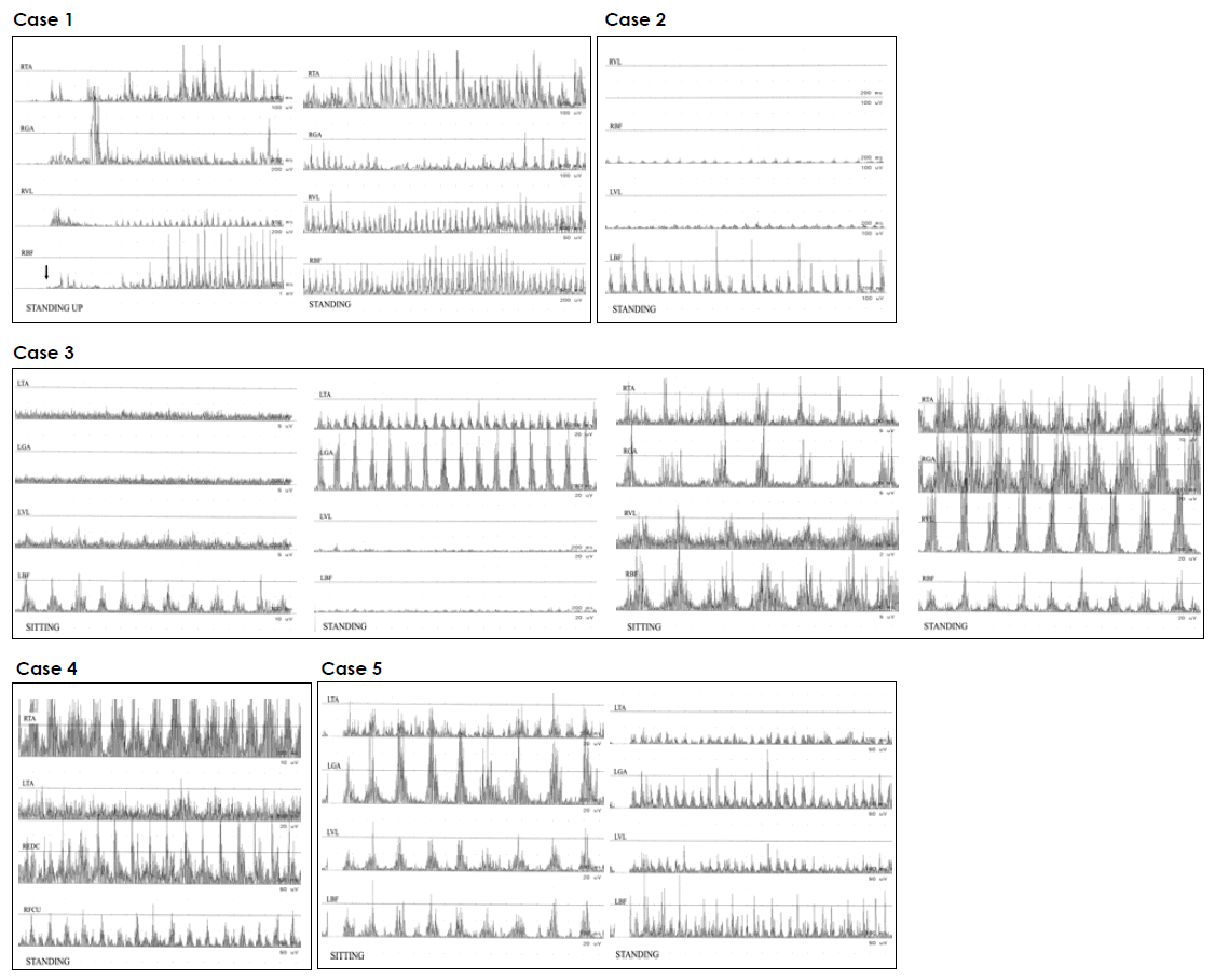
Table 1.Clinical features and EMG findings of five PD patients with leg tremor on standing
| Case no. | Orthostatic tremor | Parkinsonism | Treatment | ||||||
|---|---|---|---|---|---|---|---|---|---|
|
|
|||||||||
| Side | Frequency | Contraction pattern | Age at onset | Symptoms and signs | Age at onset | H & Y stage | Medication | Response | |
| 1 | Bilateral (right, predominant) | 6 | Alternating | 61 |
Gait disturbance Bradykinesia Rigidity, suspicious Decreased arm swing Masked face |
62 | II |
Propranolol Clonazepam* |
Good |
| 2 | Bilateral (left, predominant) | 6–7 | Alternating | 60 | Rigidity | 60 | I |
Benztropine* Propranolol* Clonazepam* Levodopa* |
Poor |
| 3 | Bilateral (right, predominant) | 4–5 |
Right, synchronous Left, alternating |
61 |
Rest tremor Rigidity Bradykinesia |
63 | II |
Benztropine* Clonazepam |
Good |
| 4 | Right | 3–4 |
Distal, synchronous Proximal, alternating |
45 |
Rest tremor Rigidity Braykinesia |
45 | I |
Levodopa Propranolol Clonazepam Trihexyphenidyl |
Good |
| 5 | Left | 16–17 | Synchronous | 47 | Rest tremor | 48 | I |
Selegiline Ropinirole Benztropine |
Good |
- 1. Gerschlager W, Münchau A, Katzenschlager R, Brown P, Rothwell JC, Quinn N, et al. Natural history and syndromic associations of orthostatic tremor: a review of 41 patients. Mov Disord 2004;19:788–795.ArticlePubMed
- 2. Heilman KM. Orthostatic tremor. Arch Neurol 1984;41:880–881.ArticlePubMed
- 3. Deuschl G, Bain P, Brin M. Consensus statement of the movement disorder society on tremor. Ad hoc scientific committee. Mov Disord 1998;13(Suppl 3):2–23.Article
- 4. Thomas A, Bonanni L, Antonini A, Barone P, Onofrj M. Dopa-responsive pseudo-orthostatic tremor in parkinsonism. Mov Disord 2007;22:1652–1656.ArticlePubMed
- 5. Langston JW, Widner H, Goetz CG, Brooks D, Fahn S, Freeman T, et al. Core assessment program for intracerebral transplantations (CAPIT). Mov Disord 1992;7:2–13.ArticlePubMed
- 6. Kim JS, Lee MC. Leg tremor mimicking orthostatic tremor as an initial manifestation of Parkinson’s disease. Mov Disord 1993;8:397–398.ArticlePubMed
- 7. Wills AJ, Thompson PD, Findley LJ, Brooks DJ. A positron emission tomography study of primary orthostatic tremor. Neurology 1996;46:747–752.ArticlePubMed
- 8. Apartis E, Tison F, Arné P, Jedynak CP, Vidailhet M. Fast orthostatic tremor in Parkinson’s disease mimicking primary orthostatic tremor. Mov Disord 2001;16:1133–1136.ArticlePubMed
- 9. Mastain B, Cassim F, Guieu J, Destee A. “Secondary” Orthostatic tremor in idiopathic parkinson’s disease. Mov Disord 1998;13(suppl 3):144.
- 10. Jankovic J, Kapadia AS. Functional decline in Parkinson disease. Arch Neurol 2001;58:1611–1615.ArticlePubMed
REFERENCES
Figure & Data
References
Citations
Citations to this article as recorded by 
- Orthostatic tremor as initial presentation of Parkinson’s disease
Y R Chiew
QJM: An International Journal of Medicine.2023; 116(7): 549. CrossRef - The ‘Postural Rhythm’ of the Ground Reaction Force during Upright Stance and Its Conversion to Body Sway—The Effect of Vision, Support Surface and Adaptation to Repeated Trials
Stefania Sozzi, Shashank Ghai, Marco Schieppati
Brain Sciences.2023; 13(7): 978. CrossRef - An unusual initial presentation of Parkinson’s disease: unilateral standing leg tremor
Jasem Yousef Al-Hashel, Walaa Ahmed Kamel, Philippe Damier, Ahmad Jasem Abdulsalam
Acta Neurologica Belgica.2020; 120(2): 415. CrossRef
Comments on this article
 PubReader
 ePub Link
 Cite
Cite
